Total & Partial Knee Arthroplasty
Knee Replacement Surgeon in Ahmedabad, Gujarat
When knee arthritis makes climbing stairs, walking in the park or even getting up from a chair painful, knee replacement can give you back years of easy movement. Dr. Hiren Patel, a knee replacement surgeon in Ahmedabad with 13+ years of experience and 11,000+ successful joint surgeries, performs total and partial knee replacement using modern implants and TriNova robotic-assisted planning for accurate alignment.

- Total, partial and revision knee replacement
- TriNova robotic and computer-navigated alignment
- High-flexion implants for Indian floor-sitting lifestyle
- Walk the same day, discharge in 2 – 3 days
What is knee replacement?
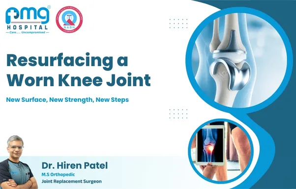
Knee replacement (knee arthroplasty) is surgery that resurfaces a knee joint worn down by arthritis or injury. Damaged cartilage and bone are removed and replaced with smooth metal and medical-grade plastic components that recreate a pain-free, moving joint. The aim is simple: remove the source of the pain and give you back a knee that does not need to be planned around - walking, stairs and getting up from a chair stop being the day's biggest decisions.
It helps to know what is not removed. The whole knee is not taken out and a metal hinge is not dropped in its place. Only about eight to ten millimetres of the worn surface is trimmed from the end of the thigh bone and the top of the shin bone - roughly the thickness of the cartilage and bone that arthritis has already destroyed - and thin metal caps are fitted over what remains. Your own ligaments, muscles, kneecap tendon and bone hold the knee together afterwards, exactly as they did before. That is why the operation is better described as resurfacing than replacement.
In Ahmedabad and across Gujarat, the patients who come to this clinic are usually between fifty-five and seventy-five, have lived with knee pain for five to ten years, and have already tried medicines, physiotherapy and often two or three rounds of injections. The decision to operate is almost never made on the first visit. It is made when the knee has stopped responding to everything else and has started shrinking the size of your day.
Who needs knee replacement?
Knee replacement is recommended when the cartilage is worn down and non-surgical treatments no longer control pain. Typical patients have trouble walking more than a few minutes, night pain, bow-legged deformity and swelling that returns quickly.
- Severe osteoarthritis or rheumatoid arthritis
- Knee pain despite medicines, injections and physiotherapy
- Difficulty climbing stairs, squatting or sitting cross-legged
- Deformity such as bowing or knock-knee
- Failed previous knee surgery or loosened implant
- Post-traumatic arthritis after an old fracture or ligament injury
- Avascular necrosis or collapse of the femoral condyle
Age on its own is neither a qualification nor a disqualification. A sixty-year-old with a bone-on-bone knee, a fifteen-degree bow and daily painkillers needs surgery more than an eighty-year-old who walks comfortably to the temple every morning. What is assessed is the gap between the life you want and the life the knee currently allows.
Types of knee replacement offered
Not every arthritic knee needs a total replacement. The right choice depends on which compartments are damaged and your activity goals. For patients who want the most precise option available, TriNova knee replacement combines ROSA robotic-assisted planning with a muscle-sparing Subvastus approach and tourniquet-less technique in a single protocol.
- Total knee replacement - all three compartments resurfaced
- Partial (unicondylar) knee replacement - smaller incision, faster recovery
- Revision knee replacement for failed or infected implants
- TriNova robotic-assisted knee replacement - ROSA precision technique
- Bilateral knee replacement in one sitting for selected patients
| Type | Best suited for | Hospital stay | Typical return to routine |
|---|---|---|---|
| Total knee replacement | Damage in two or three compartments, deformity, inflammatory arthritis | 2 – 3 days | 6 – 12 weeks |
| Partial knee replacement | Isolated inner (medial) compartment wear with intact ligaments | 1 – 2 days | 4 – 8 weeks |
| TriNova robotic knee replacement | Patients wanting maximum alignment accuracy and a gentler first fortnight | 2 – 3 days | 6 – 10 weeks |
| Revision knee replacement | Loosening, instability, stiffness or infection in a previous implant | 4 – 7 days | 3 – 6 months |
| Bilateral (both knees together) | Both knees severely arthritic, patient medically fit, good cardiac status | 3 – 5 days | 8 – 14 weeks |
Timelines are typical ranges for this practice and are individualised after your assessment.
Diagnosis before knee replacement
No one should be scheduled for a knee replacement on the strength of a single X-ray. A confident diagnosis is built from three things that must agree with each other: what the knee is doing to your daily life, what the examination finds, and what the imaging shows. When those three point in the same direction, the decision becomes obvious to both surgeon and patient. When they disagree, the answer is usually more investigation - not surgery.
How is knee damage diagnosed?
The consultation starts with questions that sound ordinary and matter enormously. How far can you walk before you must sit down? Does the pain wake you at night, or only appear after activity? Can you get up from a chair without pushing off with your hands? Can you still use an Indian toilet, kneel for prayer, or sit on the floor at a family function? How many painkillers do you take in a week, and has that number gone up over the past year? These answers describe the disability, and disability - not the X-ray - is what surgery treats.
The physical examination then looks for objective signs: the alignment of the leg while standing, how far the knee straightens and bends, swelling or fluid, grinding felt under the kneecap, tenderness along the joint line, stability of the collateral and cruciate ligaments, wasting of the thigh muscle measured with a tape, and your walking pattern across the room. The hip and lower spine are examined in the same visit, because a stiff arthritic hip and an irritated L3–L4 nerve root can both send pain to the knee and would not improve after a knee replacement.
What X-rays are needed?
X-rays remain the single most useful investigation in knee arthritis, provided they are taken correctly. The essential requirement is that they are taken STANDING. A knee X-ray taken lying down can look almost normal in a joint that collapses completely under body weight, because gravity is what closes the worn space. Films taken lying down are the most common reason a patient is told the arthritis is mild when it is not.
- Standing AP (front) view of both knees together, for comparison and joint space assessment
- Lateral (side) view, showing patellar height, bone spurs and any previous implant
- Skyline or Merchant view, which exposes wear behind the kneecap that other views miss
- Rosenberg 45° standing flexion view, which detects early back-of-the-knee cartilage loss
- Full-length hip-to-ankle scanogram, mandatory for robotic and computer-navigated planning
The scanogram deserves a special mention. It measures the true mechanical axis of the limb from the centre of the hip to the centre of the ankle and quantifies the deformity in degrees - a bow of six degrees and a bow of eighteen degrees are managed very differently. Arthritis severity is then graded on the Kellgren–Lawrence scale from 1 to 4; grade 4 means bone touching bone, and that grade combined with genuine disability is the strongest indication for replacement.
Is an MRI required?
For most patients with advanced arthritis, an MRI adds cost, not information. When the standing X-ray already shows bone against bone, an MRI simply confirms in expensive detail what is visible for a few hundred rupees. Many patients arrive with an MRI reporting a meniscus tear and are convinced this is the problem, when the meniscus in a severely arthritic knee is almost always degenerate - arthroscopy to trim it would not relieve their pain and could accelerate the wear.
An MRI genuinely helps in a smaller set of situations:
- Severe pain with a near-normal X-ray, where early cartilage loss is suspected
- Suspected avascular necrosis or a subchondral insufficiency fracture, especially sudden pain in an osteoporotic knee
- Ligament injury being considered alongside arthritis in a younger patient
- Assessment of a soft-tissue swelling, cyst or suspected tumour
- Planning a partial knee replacement, when the state of the other compartments and the ACL must be confirmed
What other tests are done before surgery?
Once surgery is decided, the workup shifts from the knee to the whole patient. The purpose is to make the operation safe and to remove every source of infection before an implant goes in, because a joint replacement has no blood supply of its own and cannot fight bacteria the way normal tissue can.
- Complete blood count, ESR and CRP - a baseline and a screen for hidden infection
- Fasting sugar and HbA1c - surgery is deferred if HbA1c is above roughly 7.5–8%, as infection risk rises sharply
- Kidney and liver function, serum electrolytes, coagulation profile and blood grouping
- Viral markers (HIV, HBsAg, HCV) and urine routine with culture where indicated
- Vitamin D and calcium levels, frequently low in Gujarati patients and corrected before surgery
- ECG for everyone; 2D echocardiogram, chest X-ray and physician or cardiologist clearance where age or history requires it
- Dental examination - an untreated abscess or infected tooth must be cleared first
- Screening for skin infection, fungal rash, or an active urinary infection near the surgical date
- Pre-anaesthesia consultation, where your anaesthetist plans the block and reviews all regular medicines
Medicines to discuss in advance
Blood thinners, aspirin, some diabetes medicines, hormone therapy and disease-modifying drugs used for rheumatoid arthritis usually need adjustment several days before surgery. Bring your actual strips or a written list rather than relying on memory, and never stop a cardiac medicine on your own.
How does the surgeon decide if surgery is needed?
Three conditions must be satisfied together. First, the pain must be limiting your life in ways you can describe concretely - not a vague ache, but a stopped morning walk, a bedroom moved to the ground floor, cancelled travel, or a sleep pattern broken by night pain. Second, the imaging must show structural damage that explains that pain. Third, honest non-surgical treatment must have been tried for at least three to six months and failed.
If all three are present, replacement is offered and the risks are discussed frankly. If only two are present, the plan is usually to treat what is treatable and review in six to eight weeks. Patients are never pushed; the operation works best in someone who has arrived at the decision themselves and is ready to do the physiotherapy that follows. A knee replacement done reluctantly, before the patient is convinced, tends to disappoint both sides even when it is technically perfect.
Treatment options before knee replacement
Most people with knee arthritis do not need surgery this year. A well-run non-surgical programme can give good years of relief in early and moderate arthritis, and even in advanced disease it improves the muscle strength and general fitness that make eventual surgery easier. The goal at this stage is honest: delay replacement for as long as the knee will genuinely allow, without wasting years on treatments that have already stopped working.
Medicines for knee pain
Paracetamol at an adequate dose is the sensible starting point and is safe for long-term intermittent use in most people. Anti-inflammatories such as ibuprofen, naproxen or etoricoxib work better for inflammatory flare-ups but are meant for short courses - five to ten days - not for daily consumption over years. Long-term self-medication with NSAIDs bought over the counter is one of the commonest avoidable problems seen in clinic, and it quietly damages the kidneys and stomach lining in exactly the age group most likely to be taking them.
Topical diclofenac gel gives useful relief for superficial knee pain with a fraction of the systemic exposure and is a good option for patients with hypertension, kidney disease or a history of gastric ulcer. Supplements such as glucosamine, chondroitin and diacerein are widely prescribed; the evidence for them is modest and inconsistent, and they are worth a three-month trial at most before deciding whether they actually help you. Correcting vitamin D deficiency, on the other hand, has a measurable effect on muscle strength and bone quality and is done routinely.
Physiotherapy
The quadriceps muscle is the knee's shock absorber. In an arthritic knee it wastes rapidly - pain inhibits the muscle, the muscle weakens, and the joint takes more load, which produces more pain. A structured six to twelve week programme of quadriceps and hip abductor strengthening, static contractions, straight leg raises, range-of-motion work and gait retraining breaks that cycle and can reduce pain meaningfully without any medicine at all.
Modalities such as ultrasound, TENS and hot packs are comfort measures that make exercise possible; they are not treatment on their own. Sessions in a supervised setting for the first three to four weeks, followed by a disciplined home routine, gives far better results than occasional clinic visits with nothing done in between.
Exercise and weight management
This is the single most powerful non-surgical intervention and the least popular. Every kilogram of body weight translates into roughly three to four kilograms of force across the knee during walking, and considerably more when climbing stairs. Losing five kilograms therefore removes something in the order of fifteen to twenty kilograms of load with every step - a change no injection can match.
- Aim for a 5–10% reduction in body weight; even that much produces a noticeable drop in pain
- Stationary cycling with low resistance and a high seat - the best exercise for an arthritic knee
- Swimming or pool walking, which builds fitness with almost no joint loading
- Walking on flat ground in cushioned shoes, in shorter and more frequent sessions
- Avoid deep squats, sitting cross-legged for long periods, stair-climbing as exercise, and treadmill inclines
Injections for knee pain
Corticosteroid injections are the most predictable, giving relief within a few days that typically lasts weeks to a few months. They are best used to break a severe flare or to get a patient through a wedding, a pilgrimage or an exam period. They are limited to roughly three or four in a year in the same knee because repeated steroid exposure damages cartilage, and no steroid should be given within three months of a planned replacement, as it measurably increases infection risk.
Hyaluronic acid (viscosupplementation) works best in mild to moderate arthritis where some cartilage remains. Relief builds over two to three weeks and may last six months or so. In a bone-on-bone knee it usually disappoints, and patients are told that honestly rather than sold a course of three injections that will not work.
Platelet-rich plasma (PRP) uses concentrated platelets from your own blood and is reasonable in early to moderate arthritis, particularly in younger and more active patients. The evidence is still developing and results vary between individuals. For patients medically unfit for surgery, a genicular nerve block or radiofrequency ablation can reduce pain for six to twelve months without entering the joint at all.
Lifestyle changes
Small changes at home reduce daily knee load more than most patients expect, and they matter particularly in Gujarati households where floor sitting is built into routine.
- Switch to a Western commode, or fit a raised seat over the Indian toilet
- Use a chair with armrests and a firm seat at knee height; avoid low sofas and diwans
- Sit on a chair for prayer, meals and family gatherings rather than on the floor
- Hold a walking stick in the hand opposite the painful knee - it removes about a fifth of the load
- Keep frequently used items at waist level to cut down bending and squatting
- Wear cushioned closed footwear indoors; hard tiles and thin chappals worsen morning pain
- Control diabetes, blood pressure and thyroid function - these decide how well a knee heals later
When do these treatments stop working?
There is usually a clear turning point, and patients recognise it in hindsight. Watch for these signals, which together mean the conservative phase is over:
- Pain at rest or at night, waking you from sleep, rather than only after activity
- Walking distance shrinking to a few minutes, or a walking stick becoming permanent
- Painkillers needed on most days of the week just to function
- Steroid injections needed more often than every six months, with each one working for less time
- Visible deformity increasing - the bow-leg becoming obvious to family members
- Knee locking, giving way, or the leg no longer straightening fully
- Stopping walks, temple visits, travel or social events because of the knee
When three or more of these are true, continuing to manage the knee with medicines mostly buys discomfort and side effects rather than time. Waiting further also allows the deformity and muscle wasting to worsen, which makes the eventual surgery technically harder and the recovery slower. This is the point at which knee replacement is discussed as the reasonable next step rather than the last resort.
Knee replacement surgery: step by step
Patients are far less anxious once they know exactly what will happen and in what order. Here is the sequence, from the moment you reach the hospital to the moment the dressing goes on.
What happens on the day of surgery?
Admission is usually the previous evening or early on the morning of surgery. You will have been asked to stop eating six to eight hours before and clear fluids two hours before. A bath with an antiseptic solution is done the night before and again in the morning; hair around the surgical area is clipped, not shaved, immediately before the procedure, because shaving causes micro-cuts that harbour bacteria.
On the ward you will change into a hospital gown, jewellery and dentures are removed, an intravenous line is started and the first dose of antibiotic is given within an hour of the incision. The surgeon marks the correct leg with a marker pen in your presence and you confirm it - a step that is never skipped. Compression stockings go on, consent is completed, and you are wheeled to the operation theatre, where the whole team runs a final safety checklist confirming your name, the procedure, the side and the implants kept ready.
How is anaesthesia given?
Most knee replacements here are done under spinal anaesthesia. A fine needle places local anaesthetic in the lower back, and within minutes the legs become heavy and completely numb while you remain awake and breathing on your own. Spinal anaesthesia is associated with less blood loss, less nausea and a clearer head afterwards than general anaesthesia, and it is the preferred technique for most older patients.
It is combined with an adductor canal block - a nerve block placed under ultrasound guidance in the thigh that numbs the pain fibres of the knee while sparing the quadriceps muscle, so you can still lift your leg and walk the same evening. Light sedation is added so that most patients simply sleep through the procedure. General anaesthesia is used when a spinal is not possible, for example with certain spine problems or blood thinner use. Throughout, your heart rate, blood pressure and oxygen levels are monitored continuously.
How is the damaged bone removed?
A midline incision of roughly ten to fifteen centimetres is made over the front of the knee. In the Subvastus approach used in the TriNova protocol, the surgeon works underneath the vastus medialis muscle rather than cutting through the quadriceps tendon, which preserves the muscle that lifts your leg - one of the main reasons straight-leg raising and walking come back faster.
The kneecap is moved aside, the joint is exposed, and osteophytes - the sharp bone spurs that block movement and tent the ligaments - are removed. Then the resurfacing cuts are made: a few precise millimetres from the end of the femur and the top of the tibia, at angles calculated from your scanogram. In conventional surgery these are guided by mechanical jigs. With ROSA robotic assistance, the arm holds the cutting guide at the planned position and the screen displays the actual angles and gaps in real time, so any deviation is corrected before a single cut is made. The amount of bone removed corresponds to the thickness of the implant that will replace it - nothing extra.
How is the artificial joint placed?
Trial components are fitted first. This is the most important part of the operation and the least visible on any video: the surgeon checks that the knee straightens fully, bends deeply, and that the gaps between the bones are equal and balanced on the inner and outer sides both when straight and when bent. Ligaments are released in small, measured steps until the balance is right. An implant that is perfectly positioned but poorly balanced will feel unstable or tight; the numbers on the robotic screen guide this, but the surgeon's judgement decides it.
Once the trial feels correct, the bone surfaces are cleaned with pulsed lavage and the definitive components are fixed with antibiotic-loaded bone cement, which sets in about ten minutes. The polyethylene insert is locked into the tibial tray, and the back of the kneecap is either resurfaced with a plastic button or smoothed and denervated, depending on how worn it is. Tranexamic acid is given to reduce blood loss, and the tourniquet-less technique means the thigh is never squeezed by a pressure cuff - a small detail that noticeably reduces thigh pain and swelling in the first week.
How is the knee tested?
Before closing, the knee is put through a complete examination on the table. It must straighten completely with no springy block, bend past 120 to 130 degrees, and feel stable when stressed inwards and outwards in both full extension and mid-flexion. The kneecap must glide down the centre of its groove without the surgeon holding it - the "no-thumb test". Final alignment is confirmed, and with robotic assistance the achieved angles are compared against the plan and recorded.
How is the wound closed?
A cocktail of local anaesthetic, an anti-inflammatory and adrenaline is injected into the tissues around the joint before closure - this is a major reason the first night is comfortable. The layers are then closed in sequence with absorbable sutures, and the skin is closed with a subcuticular stitch that leaves a thin line rather than railway-track marks. A waterproof, breathable dressing is applied so you can shower without covering the knee in plastic. Drains are usually unnecessary when tranexamic acid has been used.
How long does knee replacement surgery take?
Single knee, skin to skin
45 – 90 minutes
Robotic-assisted cases add roughly 10 minutes of registration and planning.
Total theatre time
About 2 hours
Includes anaesthesia, positioning, dressing and shifting.
Both knees in one sitting
2 – 2.5 hours
Offered to medically fit patients with cardiac clearance.
Families waiting outside should plan for roughly two to three hours between the patient leaving the room and returning to it. The surgeon speaks to the family immediately after the procedure, before the patient shifts out of recovery.
Knee replacement implants
Patients are often shown a price list of implants and asked to choose, which is the wrong way round. The implant should be selected by the surgeon to suit your anatomy, bone quality, deformity and lifestyle - and then explained to you, with the cost difference made clear.
What is a knee implant?
A knee implant is not a hinge or a mechanical joint. It is a set of three or four thin components that cap the worn surfaces: a curved metal shell over the end of the thigh bone (femoral component), a flat metal tray on the top of the shin bone (tibial baseplate), a smooth plastic insert that snaps into that tray and acts as the new cartilage, and sometimes a small plastic button behind the kneecap. Your own ligaments provide the stability; the implant only provides the smooth, low-friction surfaces.
What are knee implants made of?
- Cobalt-chromium-molybdenum alloy for the femoral component - hard, highly polished and extremely wear-resistant
- Titanium alloy for many tibial trays, chosen for its strength and ability to bond with bone
- Oxidised zirconium (Oxinium) as a premium ceramicised metal surface, useful for younger patients and those with metal sensitivity
- Highly cross-linked, and increasingly vitamin-E infused, ultra-high molecular weight polyethylene for the insert - this is the component that determines long-term wear
- Antibiotic-loaded PMMA bone cement to fix the components and deliver antibiotic locally
- Nickel-free or titanium-nitride coated options for documented metal allergy
Types of knee implants
| Implant design | What it does | Typically chosen for |
|---|---|---|
| Cruciate retaining (CR) | Keeps your posterior cruciate ligament | Good bone stock, mild deformity, intact PCL |
| Posterior stabilised (PS) | A post-and-cam mechanism substitutes for the removed PCL | Larger deformity, inflammatory arthritis, previous surgery |
| High-flexion | Geometry designed to allow deeper safe bending | Patients who need floor sitting or cross-legged positions |
| Medial pivot / ultra-congruent | Reproduces a more natural rotational movement | Patients seeking a more natural feel and stability |
| Unicondylar (partial) | Resurfaces only the worn compartment | Isolated medial wear with intact ACL and no deformity |
| Constrained / hinged | Provides stability the ligaments can no longer give | Revision surgery, severe deformity, ligament deficiency |
| Cementless | Bone grows directly into a coated surface | Younger patients with excellent bone quality |
How is the right implant selected?
The choice is made from your standing X-rays, the scanogram, bone quality, the magnitude of the deformity, the state of the ligaments and - importantly - how you actually live. A seventy-year-old who needs a stable, comfortable knee for temple walks has different requirements from a fifty-six year old teacher who must sit cross-legged and climb three floors daily.
Costliest is not always correct
A more expensive implant is not automatically a better one for your knee. What predicts a good result over twenty years is accurate alignment, balanced ligaments, a well-sized component and a clean, infection-free procedure. A premium implant placed a few degrees off will do worse than a standard implant placed correctly. You will always be told which features you are paying extra for and what they realistically buy you.
How long do knee implants last?
With current cross-linked polyethylene, national joint registries report that roughly ninety to ninety-five per cent of total knee replacements are still functioning at fifteen years, and more than eighty per cent at twenty to twenty-five years. Partial knee replacements have a slightly higher revision rate, but a partial that fails can usually be converted to a straightforward total replacement, which is part of why it remains attractive in younger patients.
What shortens implant life is predictable: excess body weight, high-impact activity such as running and jumping, poor alignment, deep repetitive squatting, uncontrolled diabetes and, above all, infection. What extends it is equally predictable - weight control, strong quadriceps, sensible activity, and dealing promptly with any dental, urinary or skin infection so bacteria never reach the joint through the bloodstream.
Recovery after knee replacement
Most patients stand and take a few steps the same evening after surgery. With multimodal pain control and early mobilisation, discharge is usually on day two or three. By two weeks most patients walk indoors without a support, and by six weeks they can walk outside, climb stairs and resume driving. A structured physiotherapy programme is essential for the best result.
Here is what each stage actually looks like.
First 24 hours
Sitting up, standing and the first steps
You return to the room with the leg wrapped and elevated. Sensation comes back over four to six hours; ankle pumps and static quadriceps contractions start immediately. Sips of water are allowed at two hours and light food at four to six hours. The physiotherapist gets you standing and walking a few steps the same evening. Ice is applied for fifteen to twenty minutes several times, a urinary catheter is avoided in most patients, and the first night is usually the most disturbed one - expect broken sleep rather than severe pain.
Days 2 – 7
Walker to corridor, and going home
Walking distance increases each day, stair practice is added, and discharge happens on day two or three for a single knee. Knee bending typically reaches 70 to 90 degrees by the end of the week. Swelling peaks around day three to five and is managed with elevation, ice and ankle pumps. The dressing is inspected around day five to seven. You will go home with a walker, a written exercise sheet, pain medicines, a blood thinner and clear instructions on what is normal and what is not.
Weeks 2 – 4
Independence at home
The walker is exchanged for a stick between weeks two and three, and many patients are walking indoors unaided by the end of week two. Bending reaches 100 to 110 degrees. Light kitchen work and moving around the house become comfortable, sutures are removed or absorbed by day twelve to fourteen, and you can shower normally. Swelling in the evening after an active day is expected and settles overnight with elevation.
6 weeks
Outdoors, stairs and back to work
This is the review point where most patients say the knee has clearly turned the corner. Expect a twenty to thirty minute outdoor walk, comfortable stairs with a railing, bending of 110 to 120 degrees, no walking aid, and a return to desk work and driving. Supervised physiotherapy usually converts to a home programme at this stage.
3 months
Near-normal daily function
Walking looks and feels normal, strength is roughly seventy to eighty per cent restored, and travel, social events and longer walks are back on. Most patients report going through parts of the day without thinking about the knee at all - the real marker of a successful replacement.
6 – 12 months
Final strength and settling
Quadriceps strength continues to improve for up to a year. A patch of numbness on the outer side of the scar, mild warmth over the knee and occasional clicking are normal and gradually fade. Follow-up is at six weeks, three months, one year, and then every two to three years with an X-ray to document that the implant remains well fixed.
How long does full recovery take?
Functional recovery - walking, stairs, driving, work, normal household life - takes about three months. Complete recovery, meaning full strength, endurance, confidence on uneven ground and the disappearance of residual swelling, takes six to twelve months. Patients who arrive with strong thigh muscles, controlled sugars and a healthy weight consistently reach these milestones earlier than the averages quoted here.
Life after knee replacement
These are the questions patients actually ask, in the order they ask them. The timelines below are the usual pattern for a straightforward single knee replacement; your surgeon will adjust them for your knee, and no milestone should be forced against pain.
When can you walk after surgery?
The same day. Once the spinal wears off, usually four to six hours after surgery, the physiotherapist helps you stand and take a few steps beside the bed with a walker. This is not a stunt - early walking reduces clot risk, controls swelling and prevents the stiffness that develops if the knee stays still. By the second day most patients walk to the bathroom and along the corridor.
When can you climb stairs?
Stair practice starts before discharge, usually on day two or three, and the rule is simple: good leg up first, operated leg down first, hand always on the railing. In the first fortnight, stairs are climbed once or twice a day rather than repeatedly. Comfortable, unassisted stair climbing typically returns between four and six weeks.
When can you drive?
For a right knee, four to six weeks; for a left knee in an automatic car, three to four weeks. Two conditions are non-negotiable: you must be off strong painkillers, and you must be able to slam the brake without hesitation. Test this in an empty parking area before taking on Ahmedabad traffic. As a passenger you can travel by car within a few days, sitting in the front seat pushed back with the leg extended.
When can you return to work?
- Desk or office work: 3 – 4 weeks, with breaks to stand and walk every hour
- Work from home or light supervisory work: often from the second week
- Teaching, retail or work involving standing for hours: 8 – 12 weeks
- Frequent travel, site visits or field work: 8 – 12 weeks
- Heavy manual labour, ladder work or squatting jobs: 3 – 4 months, sometimes with a change of duties
When can you exercise?
Walking is exercise from day one and remains the backbone of recovery. Stationary cycling begins as soon as the knee bends past about 100 degrees, usually in week three, starting with partial revolutions backwards and forwards. Swimming and pool walking are excellent once the wound is fully healed, at about four to six weeks. Light gym work - leg press within a limited arc, resistance bands, upper body training - can restart at six to eight weeks under guidance. Yoga is fine with modifications, avoiding deep kneeling postures.
When can you travel?
Short car journeys are comfortable from two to three weeks, stopping every hour to stand and stretch. Longer domestic travel and flights are advised after four to six weeks, with compression stockings, aisle seating, ankle pumps every half hour and good hydration to protect against clots. Pilgrimage travel that involves long walks, steps and floor sitting is best deferred to three months. Modern implants do sometimes trigger airport metal detectors; a card from the clinic makes security simple.
What activities should you avoid?
- Running, jogging, jumping and high-impact aerobics - the fastest way to wear the plastic insert
- Contact and twisting sports such as kabaddi, football and singles tennis
- Deep squatting and kneeling directly on a hard floor
- Indian-style toilets, permanently
- Lifting weights above roughly 15 – 20 kg, especially while turning
- Sitting cross-legged for long stretches, even if your implant allows it briefly
- Climbing on stools or ladders, where a fall risks a fracture around the implant
- Ignoring any dental, urinary or skin infection - treat it promptly and mention your implant to the treating doctor
Physiotherapy after knee replacement
Why is physiotherapy important?
The surgery gives you a painless joint. Physiotherapy is what turns that joint into a working knee. Two things are being fought in the first six weeks: scar tissue, which forms in the knee and becomes progressively harder to stretch, and quadriceps inhibition, in which the thigh muscle refuses to fire properly after surgery. Both respond well to daily work in the early window and poorly to catching up later. Stiffness that sets in by six to eight weeks may need a manipulation under anaesthesia to correct - an avoidable setback in almost every case.
Exercises after knee replacement
The early programme is short, frequent and unglamorous - three to four sessions a day of ten to fifteen minutes beats one long session.
- Ankle pumps - 10 repetitions every hour while awake, from the day of surgery, to protect against clots
- Static quadriceps - press the back of the knee down into the bed, hold 5 seconds, 10 repetitions
- Static hamstrings - press the heel down into the bed, hold 5 seconds
- Straight leg raise - lift the leg 20 to 30 cm keeping the knee locked; the single best indicator of quadriceps recovery
- Knee extension over a rolled towel under the ankle - the exercise that prevents a permanent bent knee, and the one most often skipped
- Short arc quadriceps with a bolster under the knee
- Hip abduction, sliding the leg out to the side while lying flat
The one exercise that matters most
Full straightening. A knee that bends beautifully but cannot straighten completely will limp, tire quickly and ache in the back and the other knee. Spend ten minutes twice a day lying flat with a rolled towel under the ankle and nothing under the knee, letting gravity push it straight.
Knee bending exercises
Bending is regained through repetition, not force. Heel slides in bed, seated knee bends with the foot drawn back under the chair, gravity-assisted dangling over the edge of the bed with a gentle pull from the other leg, wall slides, and - from about week three - a stationary cycle, first rocking back and forth and then completing full revolutions.
| Stage | Bending target | How it should feel |
|---|---|---|
| Day 3 – 7 | 70° – 90° | Tight and stretching; not sharp |
| Week 2 – 3 | 100° – 110° | Easier each day, sore for an hour afterwards |
| Week 4 – 6 | 110° – 120° | Comfortable enough to cycle and use stairs |
| Month 3 | 120° – 130° | Only end-range stretch is felt |
Targets are typical; the surgeon sets your individual goal based on how much the knee bent before surgery.
Walking exercises
Walk with a heel-first, toe-off pattern and a fully straight knee at the moment the heel lands. Short frequent walks build the pattern better than one long walk: five minutes several times a day in week one, ten to fifteen minutes three times a day by week three, and twenty to thirty minutes twice a day by six weeks. The walker is discarded when you stop leaning on it, and the stick when you can walk ten minutes without limping - not on a fixed calendar date.
Strengthening exercises
From weeks three to four, once the wound has healed and swelling is controlled, resistance is added: mini squats limited to thirty or forty degrees while holding a support, sit-to-stand repetitions from a firm chair, step-ups on a low step, resistance band work for the hip and thigh, calf raises, stationary cycling with light resistance and, later, a leg press through a limited arc. Hip and core strength are trained alongside, because a weak hip makes the knee work harder with every step.
How long is physiotherapy needed?
Supervised sessions typically run for four to six weeks - daily in hospital, then two or three times a week. A structured home programme continues for three to six months, and a short maintenance routine of fifteen to twenty minutes should become permanent. The knee replacement lasts decades; the quadriceps muscle protecting it needs re-earning every few weeks. Patients who stop exercising at three months are the ones who return at two years complaining that the knee has "become weak".
Pain after knee replacement
Is knee replacement surgery painful?
The surgery itself is completely painless - the leg is numb from the spinal anaesthetic and nerve block, and most patients are lightly sedated and sleep through it. The honest answer about afterwards is that the first forty-eight to seventy-two hours are uncomfortable rather than severe, and are well controlled with modern protocols. Patients consistently describe it as a deep ache and tightness with sharper pain during physiotherapy, not the constant agony they had been warned about by relatives who had surgery fifteen or twenty years ago. Pain relief after joint replacement has changed more in the last decade than almost any other part of the operation.
How is pain controlled after surgery?
Nothing here relies on a single strong painkiller. Several small measures are layered so that each one can be kept at a low, safe dose - this is what multimodal analgesia means in practice.
- Adductor canal nerve block placed under ultrasound before surgery, numbing pain fibres while preserving muscle power
- Local infiltration of an anaesthetic cocktail into the tissues around the joint before closure
- Scheduled intravenous paracetamol and an anti-inflammatory, given by the clock rather than on demand
- Opioids reserved as rescue medication only, in small doses, to avoid nausea and drowsiness
- Tranexamic acid to reduce bleeding into the joint - less blood inside the knee means less swelling and less pain
- Tourniquet-less surgery, which avoids the thigh pain and muscle bruising caused by a pressure cuff
- Ice and elevation for the first seventy-two hours, and compression to control swelling
- Early walking, which sounds counter-intuitive but genuinely reduces pain from the second day onwards
How long does pain last?
| Period | What to expect |
|---|---|
| Days 1 – 3 | Sharpest phase. Deep ache and tightness, controlled by scheduled medicines and ice. |
| Days 4 – 14 | Steadily improving. Pain now mostly during exercise and at the end of an active day. |
| Weeks 3 – 6 | Discomfort rather than pain, mainly with stairs and physiotherapy. Night pain should have gone. |
| Weeks 6 – 12 | Occasional pulling or tightness at the end range of bending. Regular painkillers no longer needed. |
| 3 – 12 months | Mild swelling or warmth after a long day, and numbness beside the scar, both of which fade. |
A useful benchmark: by six weeks, the great majority of patients report that day-to-day pain is already less than the arthritis pain they lived with before surgery.
When is pain after surgery not normal?
Recovery pain follows a downward trend. Anything that reverses that trend deserves a phone call the same day rather than a wait until the next appointment.
Contact the clinic immediately if you notice
- Pain that is increasing rather than settling after the fifth day
- Fever above 100.4°F, chills, or feeling generally unwell
- Wound discharge, spreading redness, or a wound that stays wet beyond a few days
- Calf pain, tenderness or swelling, especially with breathlessness - possible clot
- Sudden inability to bear weight, or a knee that gives way
- Pain not relieved by your prescribed medicines at all
- Pain that returns weeks or months after a completely pain-free period
Late infection is uncommon but is the reason patients are told to treat dental abscesses, urinary infections and skin infections promptly for the rest of their lives, and to mention the implant to any doctor treating them. Caught early, an infected knee replacement is usually salvageable; caught late, it often is not.
Knee replacement cost in Ahmedabad, Gujarat
Knee replacement in Ahmedabad typically costs between Rs 1.5 lakh and Rs 4 lakh per knee, depending on implant type, whether TriNova robotic assistance is used, and hospital room category. The clinic provides a written, itemised estimate covering the implant, surgeon fees, hospital stay and physiotherapy before you decide, and cashless pre-authorisation is coordinated with major insurers and TPAs.
Knee replacement patients travel to this Ahmedabad centre from across Gujarat - Gandhinagar, Nadiad, Mehsana, Anand, Surat, Vadodara and Rajkot - with pre-operative workup and admission planned within a short single visit.
| What the estimate covers | Included |
|---|---|
| Implant and instrumentation | Component cost by design and material, stated separately |
| Surgeon and anaesthesia fees | Including the nerve block and intra-operative monitoring |
| Hospital stay | Room category, nursing, OT charges and consumables for 2 – 3 days |
| Investigations and medicines | Pre-operative workup, in-hospital medicines and dressings |
| Physiotherapy | In-hospital sessions and the initial supervised programme |
| Follow-up | Reviews at 6 weeks, 3 months and 1 year |
Figures are indicative for Ahmedabad, Gujarat and are confirmed in writing after your consultation.

13+ yrs
Experience
11,000+
Joint surgeries
Meet your surgeon
Knee Replacement Surgery Specialist in Ahmedabad, Gujarat
Dr. Hiren Patel is a knee replacement surgeon in Ahmedabad with 13+ years of practice and 11,000+ successful joint surgeries at PMG Hospital, Gota. His training includes international fellowships in joint replacement across France, Switzerland, Italy and South Korea, alongside mentorship under senior Indian arthroplasty surgeons - the background behind both the CROWN partial-replacement technique and TriNova robotic-assisted knee replacement offered here.
Consultations run Monday to Saturday at Gota, with Sunday appointments on request for patients travelling from other districts of Gujarat. Second opinions on X-rays and on a knee replacement already advised elsewhere are welcome, including for patients who ultimately decide against surgery.
Frequently asked questions
How long does a knee replacement last?
Modern implants with highly cross-linked polyethylene last well beyond fifteen years in the large majority of patients, and international joint registries report that roughly eight to nine out of ten knees are still working at twenty years. Longevity depends less on the brand printed on the box and more on body weight, the accuracy of alignment and ligament balance at the time of surgery, bone quality and whether infection is avoided. A patient who keeps weight controlled, walks daily and follows activity advice will usually outlast the numbers quoted in any brochure.
Can I sit cross-legged after knee replacement?
Many patients treated with high-flexion implants and a muscle-sparing approach do sit cross-legged for short periods after three to six months, and this is discussed openly before surgery because floor sitting matters in Gujarati households. It is allowed rather than encouraged: the knee is designed for walking, stairs and chair transfers, and repeated deep flexion increases loading on the polyethylene insert. Cross-legged sitting for prayer or a meal is reasonable; hours of squatting or sitting on the floor is not.
Is TriNova robotic knee replacement better than conventional surgery?
Robotic-assisted knee replacement improves the precision of bone cuts and gives real-time numbers for alignment and ligament balance, so the plan made on screen is what actually gets implanted. In practice this usually means a smaller error margin, less soft-tissue disturbance and a more comfortable first two weeks. Long-term survival of the implant at fifteen and twenty years is broadly similar to well-performed conventional surgery, so robotics should be understood as a tool for accuracy in the hands of an experienced arthroplasty surgeon, not as a different operation.
How much does knee replacement cost in Ahmedabad?
Knee replacement in Ahmedabad typically costs between Rs 1.5 lakh and Rs 4 lakh per knee. The variables are implant type, whether TriNova robotic assistance is used, hospital room category and the length of stay. A written, itemised estimate covering the implant, surgeon fees, hospital stay and physiotherapy is given before you decide, and cashless pre-authorisation is coordinated with major insurers and TPAs.
When can I return to work after knee replacement?
Desk work is usually resumed at three to four weeks, provided you can sit comfortably and travel safely. Work that involves standing for long hours, site visits or frequent travel takes eight to twelve weeks, and heavy manual labour or work that requires squatting is deferred to three or four months. Patients who work from home often start light work in the second week.
What tests are done before knee replacement surgery?
Standing (weight-bearing) X-rays of both knees, a full-length hip-to-ankle alignment film for robotic planning, blood counts, HbA1c and sugar profile, kidney and liver function, coagulation profile, viral markers, urine routine, ECG and, where indicated, a 2D echocardiogram and chest X-ray. A dental check and a pre-anaesthesia consultation complete the workup. An MRI is not routinely needed for advanced arthritis.
Is knee replacement surgery painful?
The surgery itself is painless because the leg is completely numb under spinal anaesthesia with a nerve block. Afterwards, the honest answer is that the first two to three days are uncomfortable rather than severe, and the pain is controlled with a multimodal protocol of local infiltration, scheduled non-opioid medicines, ice and early walking. Most patients say by the sixth week that the day-to-day pain is already less than the arthritis pain they lived with before surgery.
How long is physiotherapy needed after knee replacement?
Supervised physiotherapy runs for about four to six weeks, followed by a home programme for three to six months. The first six weeks matter most, because that is the window in which knee bending and full straightening are regained; scar tissue becomes far harder to stretch after it. A short maintenance routine of fifteen to twenty minutes continues lifelong to protect quadriceps strength.
When can I drive after knee replacement?
For a right knee replacement, driving is usually safe at four to six weeks; for a left knee in an automatic car, three to four weeks is common. Two conditions must be met first: you must be off strong painkillers, and you must be able to perform an emergency brake without hesitation. A short trial in an empty parking area before driving in Ahmedabad traffic is sensible.
What warning signs after knee replacement need immediate attention?
Pain that is increasing rather than settling after the fifth day, fever above 100.4°F, wound discharge or spreading redness, calf pain with swelling, sudden inability to bear weight, or pain that returns weeks after a pain-free period. Any of these warrants a call to the clinic on the same day rather than waiting for the next scheduled follow-up.
Still deciding about knee replacement?
Bring your standing X-rays and your current medicines to the clinic in Gota. You will be told plainly whether the knee needs surgery now, can wait, or should be managed differently - and if it can wait, what to do in the meantime.
This page is written for general education about knee replacement and is not a substitute for a consultation. Treatment, implant choice, timelines and costs are decided individually after clinical examination and imaging.